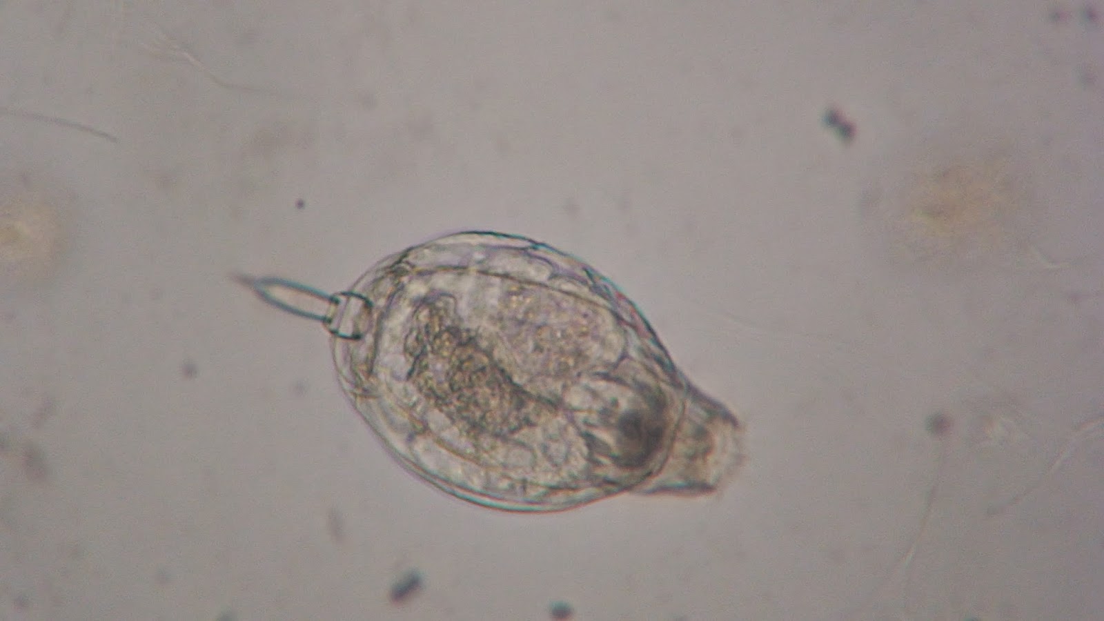
The strangest part of my final observation was the disappearance of almost all of the rotifers that once were the second most abundant organism in my aquarium. I looked almost all over for it and could not find any, except for one that was moving very slowly and did not look like it was going to make it very much longer. This is strange because the living conditions are perfect for rotifers because it is still waters and they are also able to live in areas that are moist, so having the dirt at the bottom and the plants inside the microaquarium would be a very good place for them to live (Introduction to the Rotifera, 2012) I believe the food pellet may have been gone so they started to die out. However, the perimysium were still all there and still seemed to be thriving. This makes my theory about the food pellet being gone seem less likely. Other than that I cannot think of a reason why the rotifer are all dying out.
I also found more actinophrys randomly throughout my microaquarium which is interesting because I did not see them until later on and they seem to be multiplying every observation that I make. I believe this is because they feed on other organisms and if the food pellet was all eaten, they still had food by consuming some of the other organisms. This also could be why many of the rotifers are gone if other organisms are killing them off.
From what I could find, the stentors remained almost the same and they were still relatively in the same locations as they were when I first found them. They did not seem like they had changed from earlier observations. But, the limnias that I had found in an earlier observation was nowhere to be found in this past observation. This was very fascinating considering they are very similar to the stentors, but the stentors survived and the limnias was gone (Fresh Water Invertebrates of the United States, 1989).
Bibliography:
Pennak, Robert W. Fresh-water Invertebrates of the United States: Protozoa to Mollusca. New York [etc.: John Wiley, 1989. Print.
Baqai, Aisha, Vivek Guruswanny, Janie Liu, and Gizem Rizki. "Introduction to the Rotifera." Introduction to the Rotifera. N.p., n.d. Web. 21 Nov. 2013.
Botany 111 Blog
Thursday, November 21, 2013
Sunday, November 17, 2013
11/17/13
In this past observation I noticed a very large amount of perimysium showed up and they were in all different areas of the aquarium. They tended to group together and stay in certain areas. Along with the excess perimysium that appeared there was a single midge swimming around the open area of the aquarium. It was moving forward and backward equally as much. It would also occasionally wag back and forth somewhat violently and I could not find a reason for it doing so. This midge was larger than others that I have seen in people's aquariums and it was even possible to see it without the microscope, just looking at it with the naked eye. There were also a number of actinophrys that showed up. They were mostly in the open areas, which is different than the last observation where it was in the dirt on the bottom.
A very interesting thing that I saw was a rotifer that had pinched a limnias on its tube and would not let go. It was hard to identify what was actually happening because the view of the limnias that we had was from directly above and it was somewhat hard to see (Fresh Water Invertebrates of the United States, 1989). Another unusual thing I saw was a stentor in the middle of the open area, which is unusual because it usually attaches onto the side or to dirt. And the last new organisms were volvoxes. They are very much like the actinophrys, but I could see the shorter flagellates and the daughter colonies that are part of volvox.
Bibliography:
Pennak, Robert W. Fresh-water Invertebrates of the United States: Protozoa to Mollusca. New York [etc.: John Wiley, 1989. Print.
Monday, November 11, 2013
In looking at my aquarium, there were many changes from the first time I looked. There were more organisms moving around in almost all areas of the micro aquarium.
I saw perimysiums the most out of everything in the whole aquarium. They were all moving around like slugs at a very moderate pace compared to other organisms that were around. Below is a picture of a perimysium I saw moving aroud in the middle of the aquarium where there was not much plant material or dirt.
I saw perimysiums the most out of everything in the whole aquarium. They were all moving around like slugs at a very moderate pace compared to other organisms that were around. Below is a picture of a perimysium I saw moving aroud in the middle of the aquarium where there was not much plant material or dirt.
The next most abundant organism that I found was a rotifer. They were somewhat faster than the perimysium and had more a shell type body. While viewing them I saw one standing on its two toe like flagella which was unexpected because I did not think it could balance like it was. The picture below shows the shell like body and the flagella toes that it was standing on. The toes split which cannot be seen from this image.
Another organism I observed was a stentor. The stentor was a very interesting organism to observe because it would retract back and be almost invisible. Then I could slowly see it start to make its way back out and it had little hair-like structures which would rotate around the rim of the stentor. In the image you can somewhat see the hair-like structures that are rotating around it.
Another very interesting organism in which I found in the dirt of the bottom which was not moving was an actinophrys. It is a round structure with many flagella of different sizes surrounding it. The very interesting part of the image is that it has consumed a diatom and has a bulge in it where the diatom was too big to fit in the actinophrys. You can also see the flagella surrounding it.
The last organism that I found was a gastrotrich. It was mainly moving around near what seemed to me as a cluster of food. It, like the rotifer, has a forked tail, but I never saw it use them to stand up on and it hardly moved them. It is the biggest organism that I saw in the micro aquarium and probably the fastest also.
Wednesday, October 23, 2013
Microaquarium Details
To begin setting up the microaquarium, I began with the actual microaquarium with the stand and cover and added into it the water from the water pool below the spring at Lynnhurst Cemetery. This cemetery is located off of Adair Drive in Knoxville, Tennessee. It is a spring fed pond with partial shade at N36 01.357 W83 55.731 958 ft on 10/09/2011. Then I added plants to it. These plants were Fontinalis sp. Moss. collected from the Holston River along John Sevier Highway under the I 40 Bridge. This area has partial shade exposure at N36 00.527 W83 49.549 823 ft on 10/13/2013. The second plant added was Utricularia gibba L. Flowering plant. A carnivous plant. The original material is from the south shore of Spain Lake( N 35o55 12.35" W088o20' 47.00) Camp Bella Air Road East of Sparta, Tn. in White County and grown in water tanks outside of the greenhouse at Hesler Biology Building. The University of Tennessee, Knox County, Knoxville, Tennessee on 10/13/2013.
Subscribe to:
Comments (Atom)


